Where do artifacts in MR images of the inner ear come from?
With the help of a self-constructed 3D model, senior physicians PD Dr Bela Büki and Dr Michael Bauer research together with colleagues from the Johns Hopkins University, Baltimore, USA the reasons for shadows in MR images of the inner ear and explain how the results can be useful in everyday clinical practice.
Magnetic resonance imaging is commonly used in the diagnosis of diseases of the auditory system. The inner ear is difficult to image with magnetic resonance due to its small size and structure of different tissue types. New MR techniques provide evaluable images.
3D model based on histological sections created
In MR images of the inner ear shadows typically occur. "We have provided new data that the shadows seen on magnetic resonance images show fluid movements in the inner ear due to electromagnetic activity in the magnetic field. These fluid movements are responsible for mild dizziness during an MR examination," explains Dr Bela Büki, senior physician at Krems University Hospital. Until now, little attention has been paid to these shadows in MR images.
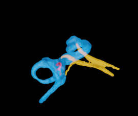
Results were received by comparing MR images of healthy people without inner ear pathology with a 3D model. The full 3D reconstruction of the inner ear was created using histological sections from a donor bone obtained from the Ear Pathology Laboratory at Harvard University, Boston, USA. The research team at KL University Hospital in Krems and Johns Hopkins University hypothesizes that certain diseases lead to altered fluid flows in the inner ear.
Anatomical structures only partly responsible for shadows
The analysis shows that "artifacts" occur predominantly in the same regions. Based on the reconstructed 3D model, it is evident that these shadows occur on the one hand because anatomical structures reduce the signal strength at these locations. On the other hand, shadows are also found at locations in the endolymph (inner ear fluids) that are not due to the presence of connective tissue structures. They are caused by movement of the fluid. The clear localization of signal disturbances in the healthy ear, as well as the realization that they are caused in part by fluid movement due to the magnetic fields of the MR equipment itself, may help to identify pathological changes in MR imaging and thus potentially contribute to improving the diagnosis of diseases of the inner ear in the future.
3D reconstruction helpful in root cause research
3D reconstruction of the human temporal bone based on histological sections has already been successfully used in causal research of neuronitis and labyrinthitis. Here, the researchers were able to show that not only viruses, but compression of the inner ear arteries can be responsible for vertigo and hearing loss (Büki et al., 2021).
Original paper


