Progress in the Personalised Treatment of Brain Tumours
Machine learning (ML) methods can quickly and accurately diagnose mutations in gliomas – primary brain tumours.
This is shown by a recent study by Karl Landsteiner University of Health Sciences (KL Krems). In this study, data from physio-metabolic magnetic resonance images were analysed to identify mutations in a metabolic gene using ML. Mutations of this gene have a significant influence on the course of the disease, and early diagnosis is important for treatment. The study also shows that there are currently still inconsistent standards for the obtaining physio-metabolic magnetic resonance images, which prevent routine clinical use of the method.
Gliomas are the most common primary brain tumours. Despite the still poor prognosis, personalised therapies can already significantly improve treatment success. However, the use of such advanced therapies is based on individual tumour data, which is not readily available for gliomas due to their location in the brain. Imaging techniques such as magnetic resonance imaging (MRI) can provide such data, but their analyses are complex, demanding and time-consuming. The Central Institute for Medical Radiology Diagnostics at St. Pölten University Hospital, a teaching and research site of KL Krems, has therefore been developing machine and deep learning methods for years in order to automate such analyses and integrate them into routine clinical operations. A further breakthrough has now been achieved.
Positive Mutation
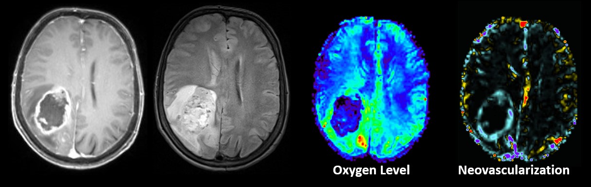
“Patients whose glioma cells carry a mutated form of the gene for isocitrate dehydrogenase (IDH) actually have better clinical prospects than those with the wild-type form,” explains Prof Andreas Stadlbauer, medical physicist at the Central Institute. “This means that the earlier we know about this mutation status, the better we can individualise treatment.” Differences in the energy metabolism of mutated and wild-type tumours help to achieve this. Thanks to earlier work by Prof Stadlbauer's team, these can be easily measured using physio-metabolic MRI, even without tissue samples. However, analysing and evaluating the data is a highly complex and time-consuming process that is difficult to integrate into clinical routine, especially as results are needed quickly due to the patients’ poor prognoses.
In the current study, the team used ML methods to analyse and interpret these data in order to obtain a result more quickly and to be able to initiate appropriate treatment steps. But how accurate are the results obtained? To assess this, the study first used data from 182 patients at University Hospital St. Pölten, whose MRI data were collected according to standardised protocols.
Positive Results
“When we saw the results of the evaluation by our ML algorithms,” explains Prof Stadlbauer, “we were very pleased. We achieved a precision of 91.7% and an accuracy of 87.5% in distinguishing between tumours with the wild-type gene and those with the mutated form. We then compared these values with ML analyses of classic clinical MRI data and were able to show that the use of physio-metabolic MRI data as a basis gave significantly better results.”
However, this superiority only held as long as the data collected in St. Pölten was analysed according to a standardised protocol. This was not the case when the ML method was applied to external data, i.e. MRI data from other hospitals' databases. In this situation, the ML method that had been trained with classic clinical MRI data proved to be more successful. “The fact that the ML analysis of physio-metabolic MRI data performed worse here is due to the fact that the technology is still young and in an experimental development phase. Data collection methods still vary from hospital to hospital, which leads to distortions in the ML analysis,” says Stadlbauer.
For the scientist, however, the problem is "only" one of standardisation, which will inevitably arise with the increasing use of physio-metabolic MRI in different hospitals. The method itself - the time-saving evaluation of physio-metabolic MRI data using ML methods - has proven to be excellent. It is therefore an excellent approach to determine the IDH mutation status of glioma patients preoperatively and to individualise treatment options.
Original Publication: Machine Learning-Based Prediction of Glioma IDH Gene Mutation Status Using Physio-Metabolic MRI of Oxygen Metabolism and Neovascularization (A Bicenter Study). A. Stadlbauer, K. Nikolic, S. Oberndorfer, F. Marhold, T. M. Kinfe, A. Meyer-Bäse, D. Alina Bistrian, O. Schnell & A. Doerfler. Cancers 2024, 16, 1102. https://doi.org/10.3390/ cancers16061102. https://kris.kl.ac.at/en/publications/machine-learning-based-prediction-of-glioma-idh-gene-mutation-sta


